
By Dr. Thomas Burnell and Dr. Bethany Turner
Next Lesson - Haemodynamic Shock
Core
The term acute coronary syndrome is used to describe a group of conditions that are caused by a sudden reduction in blood flow to the heart - this is most often caused by an atherosclerotic plaque in a coronary artery. Atherosclerotic plaques can partially occlude coronary arteries which may cause ischaemia (restriction of blood supply to tissue reducing perfusion) and further acute occlusion typically by the rupture of the plaque can cause infarction and necrosis of myocardial tissue. Acute coronary syndrome is part of ischaemic heart disease.
Acute coronary syndromes include:
- Unstable angina
- Non-ST-Elevation Myocardial Infarction (NSTEMI)
- ST-Elevation Myocardial Infarction (STEMI)
Risk factors for acute coronary syndrome can be split into two categories:
- Modifiable risk factors include: smoking, hypertension, dyslipidaemia, diabetes, obesity, sedentary behaviour.
- Non-modifiable risk factors include: male gender, advanced age, family history.
Myocardial infarctions (colloquially known as a heart attack) occur when a coronary artery is suddenly occluded, resulting in infarction (tissue death due to lack of perfusion) of the cardiac tissue that the artery supplies.
Patients will commonly present with intense, dull, central pain at rest that may radiate to the left arm, the neck and jaw. The sympathetic nervous system may be activated in response to the fall in cardiac output, resulting in nausea, generalised pallor and sweating. The patient may also be breathless (dyspnoea).
There are two main types of myocardial infarction:
- STEMI - there is complete coronary artery occlusion causing a transmural (full-thickness) injury to a region of the myocardium. The main changes on an ECG are:
- ST elevation.
- Over time the patient will develop a pathological Q wave on an ECG which will persist after recovery.
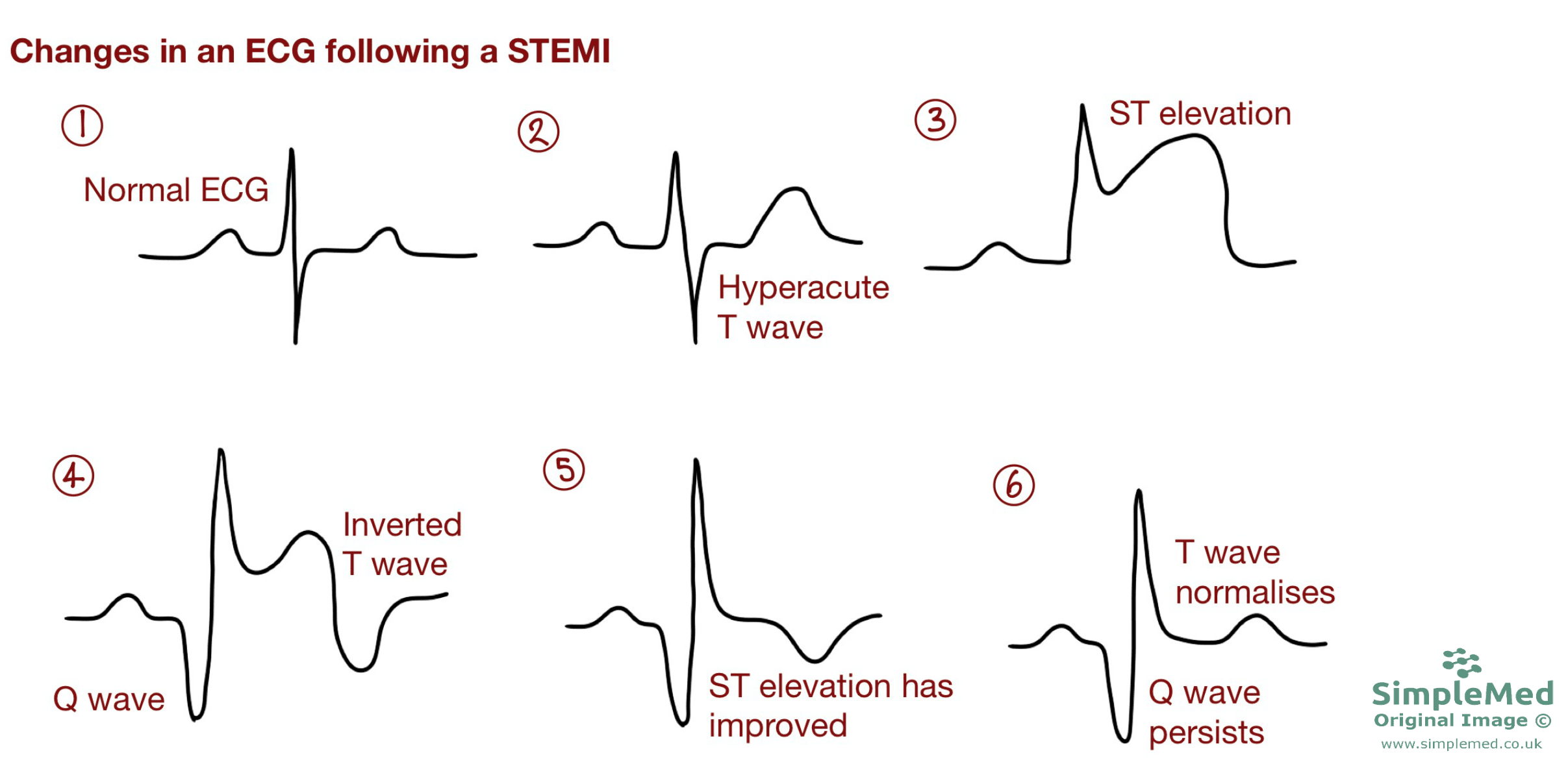
Diagram - The changes seen in the ECG of a STEMI patient over time. ST elevation is only present acutely post-MI, before disappearing. A "pathological Q wave" develops and remains permanently after a STEMI
SimpleMed original by Dr. Bethany Turner
- NSTEMI - there is partial occlusion of a coronary artery causing a sub-endocardial injury (partial-thickness - the inner layer of the heart wall is affected, while the outer layers are not fully involved). The main changes in an ECG are:
- ST segment depression and/or T-wave inversion.
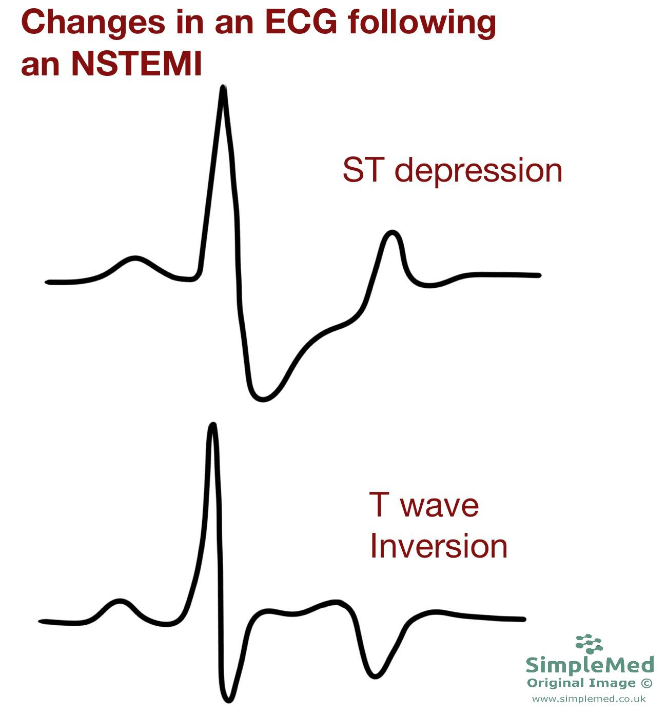
Diagram - The features of an NSTEMI on an ECG
SimpleMed Original by Dr. Bethany Turner
In an MI, the dead muscle tissue does not produce an action potential, so the ECG ‘sees through’ the infarcted area and picks up the action potential signal from the opposite side of the heart. This is directed away from the electrode and so causes a pathological Q wave. Therefore, a Q wave shows there has been muscle necrosis.
For a Q wave to be determined as pathological it must:
- Be >1 small square wide
- Be >2 small squares deep
- The depth must be more than ¼ the height of the subsequent R wave
The management of a suspected MI can be remembered by the mnemonic MONA:
- Morphine - to ease the patient's pain.
- Oxygen - only if the patient is hypoxaemic or in respiratory distress, to maintain adequate oxygen saturations.
- Nitrates - to cause vasodilation of veins to reduce cardiac return and ease strain on the heart, and to increase the blood flow through the coronary arteries.
- Aspirin - anti-platelet.
Myocardial ischaemia (angina) is when the metabolic demands of the heart are not met, causing pain. This is due to a narrowing of the lumen of the coronary arteries, typically by atherosclerosis. The plaque will be much more stable than in a STEMI or NSTEMI. This results in reduced blood flow to the myocardium. There are two main forms of myocardial ischaemia:
- Stable angina - myocardial ischaemia only occurs when the metabolic demands of the heart are increased e.g. during exercise. The pain is relieved by rest when the metabolic demand of the heart reduces again. Symptoms and signs include:
- Dull, central chest pain on exertion that is relieved by rest.
- Pain may radiate to the shoulder, jaw or left arm.
- An ECG will show ST depression in the leads corresponding to the affected area of the heart during exercise due to the ischaemia of the heart muscle.
- Unstable angina - unstable angina is cardiac pain on exertion and at rest. The pain is more intense and lasts longer than stable angina.
- Patient presents with a dull, central pain at rest which is worse on exertion.
- An ECG will show ST depression and/or T wave inversion in the leads corresponding to the affected area of myocardium.
- Unstable angina is different to NSTEMI as there is no necrosis of myocardium, therefore the troponin is normal as the tissue is intact.
Angina can be brought on by a number of causes which all result in an increased workload (and so metabolic demand) of the heart:
- Exercise - increased heart rate to meet metabolic demand.
- Stress, emotion and cold weather - all stimulate the sympathetic nervous system which leads to an increased heart rate.
- Eating - eating leads to vasodilation in the arteries to the gastrointestinal tract to aid digestion. This decreases blood pressure, which activates baroreceptors leading to an increased heart rate to restore blood pressure.
GTN can be given to relieve stable angina and may also help relieve chest pain in unstable angina, although it does not treat the underlying plaque instability. GTN causes vasodilation of veins, reducing cardiac return and therefore the workload of the heart.
- 15337